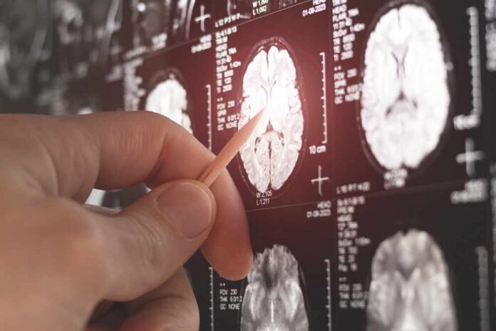
Scientists went hunting for raging brain inflammation to explain long COVID’s brain fog—and instead found a stranger, more psychological-looking signature hiding in the scans.
Story Snapshot
- New brain imaging in long COVID finds no evidence of widespread ongoing brain inflammation compared with healthy people.
- The worst long COVID symptoms track with overactive brain circuits for mood and emotion, not with a “flaming brain” picture.
- Earlier ultra–high-field scans in severe COVID did show brainstem inflammation, suggesting a time-limited, subgroup phenomenon.
- The emerging picture is heterogeneous: some inflammation early on, plus long-lasting changes in stress and emotion networks.
How The Brain-Inflammation Story Took Off
Doctors did not invent the idea of inflamed long COVID brains out of thin air. Early in the pandemic, a team from Cambridge and Oxford used ultra–high-field seven-Tesla magnetic resonance imaging to peer into the brainstem of patients hospitalized with severe COVID-19.[1] They reported abnormalities in the medulla, pons, and midbrain—key hubs for breathing, fatigue, and anxiety—that looked like neuroinflammation and correlated with longer hospital stays and worse outcomes.[1] That kind of striking, biologically tidy finding almost guarantees headlines and mechanistic enthusiasm.
Other research groups pushed even further, using positron emission tomography to track immune activity in the living brain. A Dutch team reported two long COVID patients with what looked like inflammation “throughout the brain,” with signal increases of roughly three-quarters and more than double compared with healthy volunteers.[3] For a field desperate to prove long COVID symptoms are not “all in the head,” such images looked like a smoking gun. Commentators quickly promoted a narrative: persistent brain inflammation sits at the core of long COVID.[7]
What The New Long COVID Brain Scan Study Actually Found
The newer long COVID imaging story is less dramatic and more inconvenient for simple narratives. A recent study scanning people months after infection compared long COVID patients to both healthy controls and people with multiple sclerosis, an illness well known for brain inflammation.[2] The neuroimmunology team reported no evidence of widespread brain inflammation in long COVID patients versus healthy controls, and substantially lower white matter inflammatory activity than in multiple sclerosis.[2] Objective inflammation markers looked ordinary, not “lit up.”
Researchers did see a pattern, but not the one early commentators promised. The most severe long COVID symptoms aligned with higher cellular activity in the hippocampus and amygdala—regions deeply involved in mood, memory, fear, and emotional processing.[2] Higher depression and anxiety scores, and lower quality of life, tracked with this limbic system activation.[2] That pattern suggests many long COVID sufferers may be caught in altered stress and emotion circuits rather than marching around with a uniformly inflamed brain.
Early Inflammation Versus Long-Term Brain Circuit Changes
Timing matters enormously. In the new study, people scanned within about sixteen months of infection showed higher white matter inflammatory activity than those with longer-lasting disease, hinting that inflammation may be more prominent early and then recede.[2] That would reconcile some of the contradictions: brainstem inflammation in critically ill, newly infected patients,[1] even global neuroinflammatory signals in rare, intensely symptomatic cases,[3] could be very real but time-limited. As months pass, immune fireworks fade while altered brain circuitry and subjective distress remain.
Other research supports that multi-phase picture. Laboratory scientists studying inflammatory molecules such as tumor necrosis factor alpha and interleukin-6 have found that low levels can fine-tune brain activity while higher levels suppress firing, damage cells, and trigger more inflammatory signaling.[4] That kind of dose-sensitive effect maps well onto a trajectory where an early immune surge injures vulnerable regions, and then, even after overt inflammation settles down, people live with rewired networks, fatigue, and cognitive drag.
What This Means For Patients and Politics
For patients, the message is not “nothing is wrong” but “the problem is more complex than a single inflamed-brain story.” Standard clinical magnetic resonance imaging often fails to show clear structural damage in long COVID, even in people with real and debilitating cognitive symptoms.[5] Comprehensive reviews emphasize heterogeneous findings, inconsistent correlations between scans and symptoms, and the lack of any simple imaging test that proves or rules out long COVID brain involvement.[4][8] That frustrates patients expecting a yes-or-no answer from a picture.
Scientists thought brain inflammation was driving long COVID but the scans told a different story
A new brain imaging study has found no evidence of widespread brain inflammation in patients suffering from prolonged symptoms after COVID-19 infection. Instead, the most severe…
— The Something Guy 🇿🇦 (@thesomethingguy) May 28, 2026
For public debate, these results push against both extremes. On one side, activists who insist every long COVID brain symptom must reflect ongoing global neuroinflammation now face careful data that say otherwise in many patients.[2] On the other side, commentators who hand-wave long COVID as anxiety or laziness run into measurable changes in brainstem nuclei, limbic-system activation, and molecular markers that track with cognition and quality of life.[1][2][5]
Where The Science Is Heading Next
Researchers are now mapping subtypes rather than chasing one grand unifying theory. Some groups are exploring cognitive and structural changes in long COVID cohorts that still show specific brain alterations on imaging or evidence of neurodegeneration-linked proteins.[6] Others focus on how mood, sleep, deconditioning, and chronic stress interact with lingering biological scars.[4][8] Major centers are recruiting people with long COVID brain fog into advanced imaging trials to identify which patterns correspond to which complaints and which might respond to targeted therapies.[7][8]
Sources:
[1] Web – Scientists thought brain inflammation was driving long COVID but the …
[2] Web – Brain imaging reveals changes linked to long COVID – CIDRAP
[3] Web – Using Brain Imaging to Investigate the Long-Term Changes … – CAMH
[4] Web – Ultra-powered MRI scans show damage to brain’s ‘control centre’ is …
[5] Web – Brain MRI findings in patients with post COVID-19 condition – …
[6] Web – Routine Brain MRI Findings on the Long-Term Effects of COVID-19
[7] Web – Long COVID Linked to Alzheimer’s Disease Mechanisms
[8] Web – Are you are currently experiencing brain fog from Long COVID?